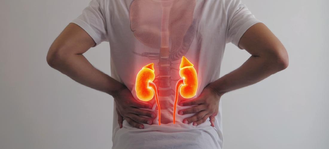
Kidney & Vascular Health
The Kidney–Blood Vessel Connection
The kidneys and the vascular system are interdependent. Every minute, the kidneys filter roughly 1.3 litres of blood. This process depends entirely on a steady, well-regulated supply through healthy arteries and veins. When those blood vessels become narrowed, blocked, or damaged, the kidneys can no longer do their job effectively. Filtration slows, waste accumulates, blood pressure climbs, and fluid balance is disrupted.
This connection works in both directions. Just as vascular disease can harm the kidneys, declining kidney function places additional strain on the cardiovascular system, raising the risk of high blood pressure, heart disease, and circulatory complications. The two systems work in tandem, which is why kidney disease and vascular disease so frequently appear side by side.
Understanding this relationship shapes how we approach care at RIVEA. Many kidney conditions that appear to be purely medical problems have an underlying vascular cause. Identifying that cause allows the possibility of targeted, minimally invasive treatments that can protect kidney function and improve outcomes. Early diagnosis matters enormously. Addressing a vascular problem before it causes lasting kidney damage is far more effective than waiting until function is significantly lost.
Common Causes
Diabetes
High blood pressure
Vascular disease affecting kidney blood supply
Genetic conditions (e.g. polycystic kidney disease)
Repeated urinary infections
Long-term use of certain medications
Common Kidney & Vascular Conditions
Chronic Kidney Disease (CKD)
Chronic kidney disease is a progressive, long-term decline in how well the kidneys function. It develops gradually, often over many years, and may go unnoticed until a significant amount of kidney function has already been lost. Because the kidneys are remarkably adaptable organs, they can continue working at a reduced capacity for a long time before symptoms become apparent, which makes routine monitoring essential for anyone at elevated risk.
How CKD progresses:
| Stage | eGFR (kidney function) | Typical symptoms | Priority |
|---|---|---|---|
| 1–2 (Early) | 60–100% | Usually none | Monitor & slow progression |
| 3 (Moderate) | 30–59% | Fatigue, mild swelling | Active management |
| 4 (Severe) | 15–29% | Swelling, nausea, breathlessness | Prepare for dialysis |
| 5 (Kidney failure) | Below 15% | Significant symptoms | Dialysis or transplant |
Why early detection matters:
- Allows lifestyle and medication changes that slow decline
- Identifies and treats underlying vascular causes
- Delays or prevents the need for dialysis
- Reduces cardiovascular risk
Renal Artery Stenosis
Renal artery stenosis is a narrowing of one or both of the arteries that supply blood to the kidneys. The most common cause is atherosclerosis or the build-up of fatty plaques within the artery wall, the same process responsible for coronary and peripheral artery disease. In younger patients, a condition called fibromuscular dysplasia can cause similar narrowing through a different mechanism.
What it causes:
- High blood pressure that is difficult to control
- Blood pressure resistant to multiple medications
- Declining kidney function over time
- Fluid retention and swelling
- In severe cases, sudden deterioration in kidney function
Renal artery stenosis vs. standard hypertension — key differences:
| Feature | Standard Hypertension | Renal Artery Stenosis |
|---|---|---|
| Response to medication | Usually responds well | Often resistant or poorly controlled |
| Onset | Gradual | May be sudden or rapidly worsening |
| Kidney function | Normal or mildly affected | Often declining |
| Treatable vascular cause | No | Yes — angioplasty/stenting |
| Imaging findings | Normal arteries | Narrowed renal artery on Doppler/CT |
How interventional radiology (IR) helps:
- Angioplasty: a small balloon opens the narrowed artery
- Stenting: a metal support keeps the artery open long-term
- Performed through a small skin puncture — no open surgery
- Can improve blood pressure control and protect kidney function
Interventional radiology offers effective, minimally invasive treatment for renal artery stenosis. Percutaneous transluminal angioplasty involves passing a small balloon-tipped catheter through the narrowed artery and inflating it to restore blood flow. In cases where the artery collapses or re-narrows, a metal stent can be placed to hold it open permanently. These procedures can meaningfully improve blood pressure control and, when performed before kidney damage becomes irreversible, help preserve long-term kidney function.
Dialysis Access Problems
For patients with advanced kidney failure, dialysis is a life-sustaining treatment but it depends entirely on reliable vascular access. Haemodialysis requires the ability to draw large volumes of blood out of the body, pass it through a filtration machine, and return it, repeatedly, during each session. Without adequate access, dialysis cannot be performed effectively.
Types of dialysis access:
- Arteriovenous (AV) fistula — surgically connected artery and vein, usually in the forearm; the preferred long-term option
- Synthetic AV graft — artificial tube connecting artery to vein when native vessels are unsuitable
- Central venous catheter — used short-term or when other access is not available
Common access problems:
| Problem | What happens | IR treatment |
|---|---|---|
| Stenosis (narrowing) | Blood flow restricted; dialysis less effective | Fistuloplasty (balloon angioplasty) |
| Thrombosis (clotting) | Access blocked; dialysis cannot proceed | Thrombectomy (clot removal) |
| Recurrent narrowing | Same site narrowing repeatedly | Stenting |
| Access failure | Access no longer usable | Access salvage procedures |
Warning signs of access problems:
- Dialysis sessions feeling less effective
- Changes in the sound or feel of the fistula
- Difficulty with needle insertion
- Swelling of the arm or hand
- Increased time to stop bleeding after needles removed
IR goals for dialysis access:
- Restore adequate blood flow without surgery
- Extend the working life of existing access
- Avoid the need for a new surgical access procedure
- Allow return to dialysis quickly — often the same day
The goal of these interventions is to extend the working life of existing access and avoid the need for surgical revision or new access creation.
Kidney Tumours
Both benign and malignant tumours can affect the kidneys. Many are now found incidentally on imaging before causing any symptoms.
Tumour types and features:
| Type | Typical behaviour | Treatment consideration |
|---|---|---|
| Renal cell carcinoma | Malignant; most common kidney cancer | Ablation or surgery depending on size/location |
| Angiomyolipoma | Usually benign; can bleed if large | Embolisation to reduce bleeding risk |
| Oncocytoma | Benign | Monitoring or biopsy to confirm |
| Complex cystic mass | May be benign or malignant | Biopsy or surgery to clarify |
Image-guided ablation — benefits at a glance:
- Destroys tumour tissue through a needle in the skin
- No open surgery or large incisions required
- Preserves healthy surrounding kidney tissue
- Particularly suitable for patients with reduced kidney function
- Suitable for patients with a single kidney
- Short hospital stay; faster recovery than surgery
Ablation techniques used:
- Radiofrequency ablation (RFA) — uses heat
- Microwave ablation (MWA) — uses heat at higher temperatures
- Cryoablation — uses extreme cold
Not every kidney tumour is suitable for ablation, and careful assessment is required to determine the most appropriate treatment for each patient. Multidisciplinary discussion informs the decision-making process. For small, localised kidney tumours in patients who are suitable candidates, image-guided ablation offers an effective alternative to surgery. These procedures are performed through a needle introduced through the skin under imaging guidance, without the need for open surgery or even a large incision. One of the important advantages of ablation is that it destroys the tumour while preserving the surrounding healthy kidney tissue, which makes it particularly valuable for patients who have reduced kidney function, a single kidney, or who are not well enough to tolerate surgery.
Renal Cysts
Renal cysts are fluid-filled sacs that develop within or on the surface of the kidney. Simple cysts are extremely common, particularly as people age, and in the majority of cases require no treatment at all. However, larger cysts, cysts that become infected, or cysts that press on surrounding structures can cause significant pain, obstruction, or recurrent urinary tract infections.
When a renal cyst may need treatment:
- Causing persistent pain in the side or back
- Pressing on the ureter, causing obstruction
- Becoming infected
- Growing significantly over time
- Showing complex internal features that raise concern
Simple vs. complex cysts:
| Feature | Simple cyst | Complex cyst |
|---|---|---|
| Contents | Clear fluid | Fluid with internal features |
| Risk of malignancy | Very low | Requires further assessment |
| Typical management | Monitor | Biopsy or treatment |
| IR treatment | Drainage ± ablation of lining | Biopsy; possible surgery |
Symptoms That May Indicate Kidney or Vascular Disease
Kidney disease can progress significantly before producing noticeable symptoms. This makes awareness of potential warning signs all the more important.
Symptoms to watch for:
| Symptom | Possible cause |
|---|---|
| Persistent swelling of feet, ankles, or face | Fluid retention from impaired kidney function |
| High blood pressure, difficult to control | Renal artery stenosis or CKD |
| Changes in urination (frequency, volume, foam) | Impaired filtration or protein in urine |
| Blood in urine | Tumour, cyst, infection, or kidney disease |
| Unexplained fatigue or weakness | Anaemia associated with kidney disease |
| Pain in the side or lower back | Cyst, obstruction, or tumour |
| Sudden decline in kidney function | Vascular event, obstruction, or acute disease |
Any of these symptoms should be evaluated by a specialist.
Higher-risk groups — seek evaluation if you have:
- Type 1 or Type 2 diabetes
- Long-standing high blood pressure
- A history of cardiovascular or peripheral vascular disease
- A family history of kidney disease
- Recurrent urinary tract infections
- Known CKD at any stage
How These Conditions Are Diagnosed
Blood tests:
- Serum creatinine — measures waste product build-up
- eGFR (Estimated Glomerular Filtration Rate) — estimates how efficiently the kidneys are filtering
- Full blood count — detects anaemia linked to kidney disease
- Electrolytes — assesses fluid and mineral balance
Urine tests:
- Protein or albumin — early marker of kidney damage
- Urine dipstick — detects blood, protein, infection
- Urine microscopy — examines cells and casts in urine
Imaging — comparison of options:
| Imaging type | What it shows | Radiation | Key uses |
|---|---|---|---|
| Ultrasound | Kidney size, cysts, obstruction | None | First-line assessment |
| Doppler ultrasound | Blood flow in renal arteries and dialysis access | None | Stenosis, access surveillance |
| CT scan | Tumours, calcification, anatomy | Yes | Detailed structural assessment |
| MRI | Soft tissue detail, vascular anatomy | None | Tumour characterisation, no radiation |
| Renal angiography | Precise arterial anatomy | Yes (X-ray) | Pre-procedure assessment; definitive vascular imaging |
Minimally Invasive Treatments
Interventional radiology has transformed the management of many kidney and vascular conditions, offering targeted treatments that achieve results comparable to surgery through much smaller incisions and with faster recovery times.
Treatment options and what they address:
| Procedure | Condition treated | How it works |
|---|---|---|
| Renal artery angioplasty | Renal artery stenosis | Balloon opens narrowed artery |
| Renal artery stenting | Renal artery stenosis | Metal support keeps artery open |
| Fistuloplasty | Dialysis access stenosis | Balloon restores blood flow |
| Thrombectomy | Dialysis access clotting | Clot mechanically or pharmacologically removed |
| Access salvage | Failing dialysis access | Combination of techniques to restore function |
| Tumour ablation (RFA/MWA/cryo) | Kidney tumours | Image-guided needle destroys tumour |
| Embolisation | Bleeding, tumour, AVM | Blood supply to target is blocked |
| Image-guided biopsy | Kidney masses | Tissue sample obtained for diagnosis |
| Cyst drainage ± ablation | Symptomatic renal cysts | Cyst drained; lining treated to prevent refilling |
IR vs. open surgery — general comparison:
| Factor | Interventional radiology | Open surgery |
|---|---|---|
| Incision | Small skin puncture | Large surgical incision |
| Anaesthesia | Local + sedation (usually) | General anaesthesia |
| Hospital stay | Same day or overnight | Several days |
| Recovery time | Days to weeks | Weeks to months |
| Risk of complications | Lower in most cases | Higher in most cases |
| Suitability | Most patients, including frail | May not suit all patients |
When to Seek Specialist Care
Consider specialist evaluation if you:
- Have been diagnosed with CKD at any stage
- Are approaching the need for dialysis
- Are already on dialysis and experiencing access problems
- Have high blood pressure resistant to medication
- Have been told you have a kidney tumour or suspicious mass
- Have renal artery narrowing identified on any scan
- Have a complex renal cyst requiring further assessment
- Have unexplained decline in kidney function
The earlier an assessment takes place, the more treatment options are available and the greater the chance of a good outcome.
Why Choose RIVEA
At RIVEA Vascular Institute, kidney and vascular conditions are assessed and treated through advanced imaging, minimally invasive procedures, and multidisciplinary collaboration.
What patients benefit from:
- Specialist expertise in vascular therapies and interventional radiology
- Advanced image-guided diagnosis and treatment
- Organ-preserving approach — protecting kidney function wherever possible
- Comprehensive dialysis access care, from assessment to salvage
- Personalised treatment planning based on individual anatomy and history
- Coordinated multidisciplinary care
- Faster recovery through minimally invasive techniques
Our goal is to protect kidney function, improve circulation, and reduce the need for major surgery wherever that is possible.