Liver Tumours: Understanding Your Diagnosis and Treatment Options
Understanding Liver Tumours
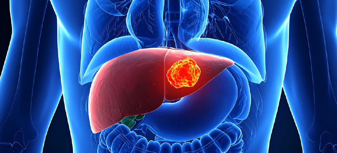
The liver is one of the body’s most resilient organs. It filters blood, supports digestion, produces vital proteins, and uniquely, has the ability to regenerate. When abnormal cells grow in the liver, they form tumours. Identifying the type and extent of tumour is the first step toward choosing the right treatment.
Primary Liver Cancer
These tumours originate in the liver. The most common type is Hepatocellular carcinoma (HCC), often seen in patients with chronic liver disease or cirrhosis. Another primary type is Cholangiocarcinoma, which arises from the bile ducts within the liver.
Secondary Liver Cancer (Liver Metastases)
These tumours begin elsewhere in the body and spread to the liver. The liver is a common site for metastases as it filters blood from much of the body. Cancers that frequently spread to the liver include colorectal, breast, lung, and pancreatic cancers.
Recognising the Symptoms
Liver tumours do not always cause noticeable symptoms in their early stages, which is why they are often detected incidentally during scans for other conditions. When symptoms do occur, they may include:
Discomfort or pain in the upper right abdomen
Unexplained weight loss or loss of appetite
Fatigue and general weakness
Jaundice (yellowing of the skin or eyes)
Abdominal swelling or a feeling of fullness
Nausea
If you are experiencing any of these symptoms, or if you have a known cancer and are being monitored for spread, speak to your doctor promptly.
Diagnosis and Assessment
Accurately diagnosing a liver tumour involves a combination of imaging and, in some cases, tissue sampling. Your medical team will build a complete picture of your condition before recommending a treatment plan.
Common diagnostic steps include:
- Blood tests – including liver function tests and tumour markers such as AFP (alpha-fetoprotein) for HCC
- Ultrasound – often the first imaging tool used to detect abnormalities in the liver
- CT scan – provides detailed cross-sectional images of the liver and surrounding structures
- MRI – particularly useful for characterising liver lesions and assessing their relationship to blood vessels
- Biopsy – a small tissue sample may be taken to confirm the tumour type, though this is not always required
Who Is Affected and Why
Liver tumours can affect a wide range of patients. There are two broad groups:
Primary liver cancer is more common in people with:
- Chronic liver disease or cirrhosis
- Hepatitis B or C infection
- Long-term alcohol use
Secondary liver tumours can develop in patients with:
- Colorectal cancer
- Breast cancer
- Lung cancer
- Pancreatic cancer
Because the liver filters blood from much of the body, it is one of the most frequent sites for cancer to metastasise.
Why Interventional Radiology (IR) for Liver Tumours?
Interventional Radiology offers a minimally invasive approach to treating liver tumours. Instead of traditional surgery requiring large incisions, IR specialists use advanced imaging guidance to navigate fine instruments through blood vessels or directly into the tumour.
Using real-time X-ray (fluoroscopy), ultrasound, or CT imaging, specialists can precisely visualize the tumour and surrounding structures, allowing treatment to be delivered exactly where it is needed while preserving healthy liver tissue.
Why the Liver Is Suited to Targeted Treatment
The liver has a unique dual blood supply and a remarkable ability to regenerate. Liver tumours, especially hepatocellular carcinoma and many metastases, derive most of their blood supply from the hepatic artery—making them especially amenable to targeted, catheter-based therapies such as embolization.
When treatment is delivered precisely to the tumour, healthy tissue can often recover and continue functioning effectively.
Interventional Radiology allows:
- Selective targeting of tumour blood vessels, sparing unaffected liver tissue
- Localized delivery of chemotherapy or radiation, reducing systemic exposure
- Image-guided ablation of small tumours, with curative intent in appropriate cases
- Treatment of tumours in surgically difficult locations
- Repeat procedures when necessary, which is often important in chronic liver disease
For patients who may not be ideal candidates for major surgery—due to tumour location, liver function, or overall health—IR provides an effective and organ-preserving alternative. In carefully selected patients, IR can be used as a definitive treatment, a bridge to transplantation, a downstaging strategy before surgery, or as part of a comprehensive multidisciplinary cancer plan. By combining precision imaging with targeted therapy, Interventional Radiology enables effective tumour control while prioritizing liver preservation and recovery.
Benefits of IR Treatments
- Minimally invasive: Small access points instead of large surgical incisions
- Faster recovery: Many patients go home the same day or after one overnight stay
- Less pain: Reduced post-procedure discomfort compared to surgery
- Targeted treatment: Delivers therapy directly to the tumour while preserving healthy tissue
- Repeatable: Can often be performed multiple times if needed
- Lower risk: Generally safer for patients who may not tolerate major surgery
For patients who may not tolerate major surgery—or whose tumours are difficult to operate on—IR can provide meaningful control with less physical strain.
RIVEA’s Core IR Treatment Approaches
At RIVEA, liver tumour treatments broadly fall into two categories: Embolization and Ablation, and when required, peripheral vascular interventions that support overall treatment safety and effectiveness.
Your care team will recommend the most appropriate strategy based on your diagnosis and overall health.
These treatments work by blocking the blood supply to liver tumours. Since tumours need nutrients from blood to grow, cutting off their supply can shrink or destroy them.
Embolization Therapies
Common techniques include:
- TACE (Transarterial Chemoembolization) – Delivers chemotherapy directly into the tumour while simultaneously blocking its blood supply
- TAE (Transarterial Embolization) – Blocks tumour blood flow without chemotherapy
- TARE / Y90 Radioembolization – Uses microscopic radioactive beads to deliver internal radiation precisely to the tumour
- Bland Embolization – Mechanical blockage of tumour-feeding vessels
These procedures are performed through a small catheter inserted typically at the wrist or groin.
Ablation Therapies
Ablation destroys tumour tissue directly using heat, cold, or electrical energy delivered through a thin probe placed into the tumour under imaging guidance.
Techniques include:
- Radiofrequency Ablation (RFA) – Heat generated by radio waves destroys tumour cells
- Microwave Ablation (MWA) – High-frequency microwaves heat and eliminate tumour tissue
- Cryoablation – Extreme cold freezes and destroys cancer cells
- Irreversible Electroporation (IRE) – Electrical pulses disrupt tumour cells while preserving nearby critical structures
Ablation is often ideal for small to medium-sized tumours and can be curative in select cases.
Peripheral Vascular Interventions
In patients with concurrent vascular disease or complex anatomy, peripheral interventions may be performed to optimise blood flow and procedural access. These can include angioplasty, stenting, or vessel management to:
- Ensure safe catheter navigation
- Improve hepatic or systemic circulation
- Reduce procedural risk
- Support overall treatment outcomes
Addressing vascular integrity is often essential in patients with underlying atherosclerosis, diabetes, or peripheral arterial disease, and forms part of a comprehensive interventional strategy.
By combining precise imaging, targeted tumour therapy, and when necessary, supportive peripheral vascular interventions, Interventional Radiology provides a comprehensive, organ-preserving approach to liver tumour management, balancing oncologic effectiveness with patient safety and recovery.
How Doctors Choose Your Treatment
Selecting the right treatment is a personalized process. Your medical team considers many factors to create a treatment plan tailored specifically to you. No two patients are exactly alike, and your treatment reflects that individuality.
Key Factors in Treatment Selection
- Tumour characteristics: Size, number, and location of tumours in your liver
- Cancer type: Whether it's primary liver cancer or spread from elsewhere
- Liver function: How well your liver is working overall
- Overall health: Your general medical condition and ability to tolerate procedures
- Previous treatments: What treatments you've already had and how you responded
- Treatment goals: Whether aiming for cure, control, or symptom relief
What to Expect
Before the Procedure
You’ll undergo blood tests and imaging scans. Medications may be adjusted temporarily. Your specialist will walk you through the procedure and answer your questions in detail.
During the Procedure
Depending on the treatment, you may receive conscious sedation or general anesthesia. Most procedures last between 1–3 hours.
After the Procedure
You’ll be monitored for several hours. Some fatigue, mild discomfort, or low-grade fever may occur for a few days. Many patients resume normal activities within a short period.
Recovery is typically significantly quicker than traditional surgery.
Is IR Treatment Right for You?
IR treatments may be an option if:
- You have liver tumours that are small to medium-sized
- Surgery is not recommended due to tumour location or your overall health
- You want a minimally invasive alternative to surgery
- You need treatment that can be repeated if necessary
- Your liver function is adequate to tolerate the procedure
However, not everyone is a candidate for IR treatments. Your medical team will carefully evaluate whether these approaches are appropriate for your specific situation.
Why RIVEA
- Dedicated Vascular & Interventional Excellence – A focused centre built around image-guided therapies
- Advanced Imaging Infrastructure – High-resolution, real-time guidance for maximum precision
- Experience in Complex Cases – Including patients not eligible for conventional surgery
- Multidisciplinary Decision-Making – Every case discussed collaboratively
- Patient-Centred Care Pathways – Clear communication, coordinated scheduling, and structured follow-up
- Commitment to Organ Preservation – Treating disease while protecting function
Click here to learn more about:
Ablation for Liver Tumours
For any inquiries, post your query here:
Ask Rivea
Contact us today to explore your options.
Call Now